Our Office
Visit Us Online

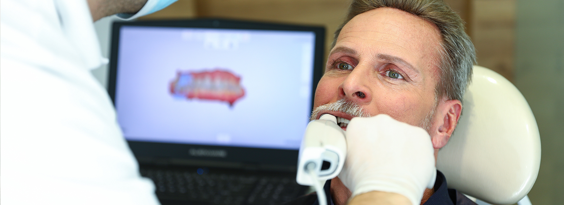
Digital impressions replace traditional putty-based trays with a handheld intraoral scanner that captures a three-dimensional map of the teeth and surrounding soft tissues. Instead of waiting for impression material to set and then shipping that physical mold to a lab, the scanner records thousands of data points and assembles them into a precise virtual model. This digital model becomes the starting point for crowns, bridges, aligners, and many restorative and orthodontic workflows.
The transition from impression tray to digital file removes many of the inconveniences patients remember from older techniques—no gagging on trays, less chairtime spent waiting for materials to harden, and no risk of distortion from handling. Clinicians gain an immediate visual reference they can review with the patient on-screen, which improves communication and helps set realistic expectations for treatment outcomes. For practices that offer same-day restorations, the digital file is the linchpin that enables rapid, predictable workflows.
Because digital impressions are standardized electronic records, they integrate cleanly into modern practice management and laboratory systems. Files can be archived, compared over time, and transmitted instantly to in-house or external dental labs. This efficiency is one reason many dental teams—particularly those offering restorative and implant care—have made digital scanning a core part of their diagnostic and treatment planning toolkit.
A typical digital scanning appointment begins with a brief explanation from the dental team about what the patient will feel and see. The intraoral wand is moved around the mouth, capturing the surface of each tooth and the contours of the gums. The process is noninvasive and generally well tolerated; because the scanner relies on light and small cameras rather than physical impression material, patients usually report minimal discomfort.
As the scanner gathers data, the software stitches individual frames together to form a continuous 3D model in real time. The clinician watches this model appear on a monitor and can immediately identify areas that need rescanning, such as moist or reflective surfaces that temporarily obscure detail. This instant feedback loop reduces the chance of remakes and ensures the file sent to the lab is complete and accurate.
For complex cases—multiple-unit bridges, implant-supported restorations, or full-arch rehabilitations—the scan may be combined with other digital records like CBCT images or digital bite registrations. The combined dataset gives the dental team a comprehensive view of hard and soft tissues, enabling more precise planning and fewer surprises during treatment.
One of the clearest advantages of digital impressions is patient comfort. Many people experience anxiety or gagging with traditional trays; digital scanning removes those physical barriers. Shorter scanning times and fewer retakes also mean less total chairtime, which contributes to a more pleasant visit overall. These practical benefits often translate into greater willingness to pursue elective and restorative treatments.
Digital impressions also improve communication. Seeing a virtual model on-screen allows clinicians to explain conditions and proposed solutions visually, which helps patients understand their treatment options and the anticipated results. Visual records can be saved and revisited during follow-up visits, providing a consistent reference point for monitoring change over time.
Another patient-centered advantage is the predictability of outcomes. Because digital files are precise and easily transferable, the restorations fabricated from them tend to require fewer adjustments at delivery. That means fewer return visits and a more seamless path from the initial consult to the final restoration.
After capture, the digital impression becomes the backbone of modern restorative and orthodontic workflows. The electronic file can be refined in specialized software, where technicians or clinicians design crowns, veneers, and aligners with exacting control over margins, contacts, and occlusion. For practices with in-house CAD/CAM milling or printing capabilities, the design can be turned into a restoration the same day.
When outside laboratories are involved, files are transmitted digitally with detailed prescription notes. This eliminates shipping delays and reduces the risk of loss or damage that accompanies physical impressions. Laboratories receiving high-quality scans can often complete their work faster, because they are working from an immediate, distortion-free representation of the patient’s anatomy.
Integration with other technologies—digital bite records, cone beam images, and implant planning software—further enhances the workflow. For implant cases, in particular, digital impressions aligned with surgical guides and radiographic data help ensure the prosthetic component matches the planned implant position, contributing to better long-term function and esthetics.
Accuracy is central to the value proposition of digital impressions. Modern scanners capture fine anatomical detail and spatial relationships with high consistency, which directly affects how well restorations fit. A precise fit reduces the need for chairside adjustments, minimizes the potential for cement gaps, and supports the longevity of crowns, bridges, and implant restorations.
Beyond single restorations, full-arch scans enable clinicians to evaluate occlusion and plan multi-unit treatments with a level of control that conventional impressions find hard to match. Digital archives also allow clinicians to compare scans taken at different times, monitoring wear, tooth movement, or soft tissue changes without additional exposure to radiographs.
Finally, digital impressions contribute to a modern standard of care by creating reproducible, verifiable records. When combined with careful clinical technique and high-quality laboratory workflows, they help deliver predictable outcomes that align with both functional needs and cosmetic goals.
Summary: Digital impressions simplify the capture of dental anatomy, enhance patient comfort, and streamline the path from diagnosis to restoration. They support faster, more accurate lab communication and enable same-day solutions when paired with in-office manufacturing. For patients in Waller, TX and nearby communities, adopting digital scans is a practical step toward predictable, comfortable dental care. If you’d like to learn more about how digital impressions are used in our practice, please contact us for additional information.
Digital impressions are three-dimensional virtual models of the teeth and surrounding tissues captured with an intraoral scanner rather than putty-based trays. The scanner records thousands of data points and the software stitches those frames into a precise digital model that can be reviewed immediately. Because the process uses light and small cameras instead of impression material, it eliminates the need for physical molds and reduces common problems like distortion and gagging.
The digital file serves as the foundation for crowns, bridges, implant restorations, and orthodontic appliances, and it can be edited or combined with other records for treatment planning. Clinicians can show the model to patients on-screen to explain conditions and proposed work, improving understanding and informed consent. Unlike conventional impressions that must be shipped, digital files are transmitted electronically, which streamlines communication with dental laboratories and manufacturing equipment.
A digital scanning appointment typically begins with a brief explanation of the procedure and what the patient will feel. The clinician moves the handheld wand around the mouth while the scanner captures the surfaces of the teeth and the contours of the gums, and most patients report little or no discomfort. The process is noninvasive and is usually faster than taking conventional impressions, with fewer pauses for material to set.
As the scan progresses, the software displays a live 3D model so the clinician can verify coverage and immediately rescan any voids or reflective areas. This instant feedback reduces the likelihood of remakes and the need for additional visits. If other records are needed, such as a digital bite registration or CBCT images, those are captured or combined to create a comprehensive dataset for planning.
Modern intraoral scanners capture fine anatomical detail and spatial relationships with high consistency, which directly influences how well restorations fit. Accurate digital data helps technicians and clinicians design margins, contacts, and occlusion with exacting control, reducing the need for chairside adjustments. When scans are performed correctly and combined with precise laboratory workflows, restorations tend to seat more predictably.
For implant cases, accuracy is enhanced when scans are aligned with radiographic data and surgical guides, ensuring the prosthetic components match the planned implant positions. Full-arch and multi-unit restorations benefit from the ability to capture continuous surfaces and evaluate occlusion digitally, providing clinicians with tools that support long-term function and esthetics. Digital archives also allow clinicians to compare scans over time, which supports monitoring and maintenance of prosthetic work.
Digital impressions integrate directly with CAD/CAM software that allows clinicians to design restorations on-screen and send milling instructions to in-office mills or 3D printers. When a practice has chairside manufacturing capabilities, the digital workflow can move from capture to design to fabrication within a single visit. This removes the need for temporary crowns and multiple appointments in many cases.
Even when outside laboratories are used, transmitting high-quality digital files eliminates shipping delays and reduces the risk of distortion associated with physical impressions. The result is a more predictable timeline and fewer remakes because lab technicians work from a distortion-free, immediate representation of the patient’s anatomy. That efficiency supports streamlined restorative care without sacrificing precision.
Yes. Digital impressions are widely used to create the accurate models needed for clear aligners, retainers, and other orthodontic appliances. The scan provides a detailed virtual record that orthodontic software uses to plan tooth movements and fabricate sequential aligner trays with controlled force application. Because the model is digital, the clinician and patient can also visualize the projected treatment stages during the consultation.
Digital records simplify communication with aligner manufacturers and allow for quick adjustments to treatment plans when necessary. They also make it easier to monitor progress by comparing scans taken at different points in therapy. These capabilities enhance both the planning and follow-up phases of orthodontic care.
Digital impression files are standardized electronic records that can be archived within a practice management system or a secure digital repository. Proper systems include encryption in transit and at rest, controlled access, and routine backups to protect patient data and ensure long-term availability. Storing scans electronically also enables clinicians to retrieve historical records for comparison and case review without reimaging the patient.
When files are sent to an outside laboratory or specialist, secure transmission protocols are used to minimize the risk of unauthorized access. Integration with practice software and laboratory portals further streamlines workflows while preserving data integrity. Practices follow applicable privacy and security guidelines to protect patient information during storage and transfer.
Most patients are suitable candidates for digital impressions, including those who have strong gag reflexes, anxiety about traditional trays, or complex restorative needs. The technology is versatile for single-unit restorations, multi-unit bridges, implant restorations, and orthodontic work, making it appropriate for a wide range of clinical situations. Because the process is noninvasive, it is generally well tolerated by children and adults alike.
There are occasional limitations, such as restricted mouth opening, heavy bleeding, or excessive saliva, which can make capturing ideal images more challenging. In complex cases clinicians may combine scans with other records like CBCT imaging or implant-level transfers to obtain the necessary detail. Ultimately the dentist evaluates each patient and tailors the imaging approach to achieve the best possible result.
Digital scanning provides immediate visual feedback that allows clinicians to correct incomplete or distorted areas during the appointment, which reduces the likelihood of retakes. Because technicians and clinicians design restorations from a precise, distortion-free model, the restorations often require fewer chairside adjustments at delivery. This efficiency contributes to smoother restorative visits and fewer follow-up appointments for refinements.
Additionally, digital workflows enable finer control over margins, contacts, and occlusion during the design phase, which helps minimize adjustments when the restoration is fitted. Combining accurate scans with careful clinical technique and high-quality lab processes supports predictable outcomes and long-term prosthetic performance. Archiving the digital records also makes it easy to reproduce or modify work if future changes are needed.
Digital impressions are safe and generally comfortable because they rely on optical scanning rather than impression materials that sit in the mouth for several minutes. The intraoral wand is lightweight and noninvasive, and most patients report little to no discomfort during capture. Because the process is faster and avoids bulky trays, it is particularly helpful for patients with gag reflex sensitivity or dental anxiety.
From an infection-control perspective, manufacturers design scanner tips and workflows to comply with standard clinical sterilization and barrier protocols. Clinicians follow established practices for cleaning and disinfecting equipment between patients to maintain a safe environment. Overall, the combination of comfort and standard safety measures makes digital scanning a patient-friendly option.
Towne Dental & Orthodontics uses digital impressions as a core tool in restorative, implant, and orthodontic workflows to improve diagnostic clarity and treatment predictability. Scans are combined with other digital records when needed to create comprehensive treatment plans, and clinicians review the virtual models with patients to explain options and expected results. Integrating digital scans into the office workflow helps coordinate care between clinicians and dental laboratories efficiently.
When in-office manufacturing is appropriate, the digital workflow supports same-day solutions by moving seamlessly from capture to design and fabrication. For lab-fabricated restorations, high-quality digital files reduce turnaround time and the potential for remakes by delivering precise, distortion-free data. This approach helps the practice deliver modern, predictable care that emphasizes patient comfort and clinical accuracy.