Our Office
Visit Us Online

Digital radiography is the contemporary approach to capturing X-ray images using electronic sensors and computer processing rather than film. This method converts X-ray energy into a digital image almost instantly, giving clinicians immediate visual feedback. For patients, that means examinations move more smoothly and decision-making can begin during the same appointment instead of waiting for film to be developed.
Unlike traditional film, which relies on chemical development and physical storage, digital images are files that can be reviewed, enhanced, and archived within a patient’s electronic record. The technology includes a variety of sensor types and imaging software, each designed to balance image clarity with patient comfort. Because images are produced and displayed digitally, teams can zoom, compare, and measure findings with tools that are not available on conventional film.
At Towne Dental & Orthodontics we integrate digital radiography into routine care to support quicker diagnoses and more efficient treatment planning. The technique is versatile—useful for routine checkups, restorative planning, orthodontic assessments, and more specialized evaluations—making it a foundational tool in modern dental practice.
One of the most significant clinical advantages of digital radiography is its ability to produce diagnostic images with lower radiation doses than traditional film X-rays. Advances in sensor sensitivity and image processing allow the system to capture clear images with less exposure, helping to minimize patient risk while preserving diagnostic value. Dosage remains low across standard dental imaging protocols, and practitioners follow established safety guidelines to protect all patients.
Lower exposure is particularly important for children, pregnant patients (when imaging is necessary and following clinical guidelines), and patients who require frequent monitoring. Because the images are produced digitally, repeated exposures for image quality checks are less common; clinicians can often adjust images on-screen to obtain necessary detail without taking additional X-rays.
Beyond dosage, digital systems make it simple to use protective practices such as shielding and precise beam collimation. When combined with contemporary protocols and well-trained clinicians, digital radiography supports a high standard of safety in everyday dental care.
Digital radiography replaces film with a compact electronic sensor that rests gently in the mouth when an image is taken. That sensor transforms X-ray photons into an electronic signal, which is then converted into a digital image by the computer. The result appears on a monitor within seconds, allowing the dentist to evaluate the image immediately and explain findings to the patient while the appointment continues.
Once captured, images are stored in the patient’s electronic chart. This storage facilitates a secure, chronological record that can be retrieved quickly for follow-up visits or treatment planning. Images can be displayed side-by-side with prior studies, enabling easy comparisons that help identify subtle changes over time—an important advantage in tracking periodontal health, cavity progression, or the stability of restorations and implants.
Digital files are also much simpler to share with other clinicians when collaboration is needed. Whether coordinating care with a specialist or transferring records for a referral, digital images can be exported in standardized formats that preserve diagnostic quality while streamlining communication.
Image enhancement tools available with digital radiography—such as contrast adjustment, magnification, and measurement overlays—give dentists more control over how they view findings. These capabilities can make it easier to spot early signs of decay, hairline fractures, bone loss, or anomalies that might be missed on lower-resolution film. The improved visualization supports more accurate diagnoses and enables clinicians to explain treatment options more clearly to patients.
Digital imaging also supports interdisciplinary planning. Orthodontists, periodontists, and oral surgeons can review the same high-quality images, often simultaneously, which helps coordinate complex treatment sequences and avoid unnecessary delays. When planning restorations or implants, clinicians can marry radiographic data with clinical records to design solutions that fit a patient’s anatomy and long-term goals.
Because the images are digital, it’s easier to document findings and treatment rationale within the chart. That documentation enhances continuity of care and can be helpful when monitoring progress or evaluating outcomes over months and years.
Digital radiography eliminates the need for hazardous chemical developers, film, and physical storage space, which reduces the environmental footprint of imaging procedures. Practices that adopt digital workflows avoid chemical waste and minimize paper usage, aligning patient care with more sustainable office operations.
From an operational perspective, immediate access to images speeds up appointment flow and reduces the administrative steps associated with film processing and archiving. Staff can retrieve images instantly, share them with colleagues electronically, and include them in patient education materials shown during the visit. This efficiency helps keep appointments on schedule and supports a clearer, more engaging conversation about oral health.
Security and privacy are also improved with robust electronic record systems. Digital images are stored within encrypted patient records and subject to the same safeguards as other protected health information. Controlled access, audit trails, and secure backups help ensure that radiographic records remain both available and confidential when they are needed for clinical care.
Digital radiography represents a meaningful upgrade from traditional film X-rays: faster results, lower radiation exposure, enhanced diagnostic tools, and environmentally friendlier workflows. The technology integrates seamlessly into general, restorative, and specialty dental care, enabling clinicians to make better-informed decisions and to communicate those decisions clearly with patients.
If you would like to learn more about how digital radiography is used in our office or what you can expect during an imaging appointment, please contact Towne Dental & Orthodontics for additional information. Our team is happy to explain the process and answer questions so you can feel confident about your care.
Digital radiography uses electronic sensors and computer processing to create X-ray images instead of photographic film. The sensor converts X-ray energy into a digital file that appears on a monitor within seconds, which lets clinicians evaluate findings immediately. This digital workflow eliminates chemical development and physical film storage while enabling on-screen tools for closer examination.
Unlike conventional film, digital images can be enhanced, magnified, and measured with software to reveal subtle details that may be harder to see otherwise. Files are stored electronically in the patient chart, making historical comparisons and retrieval much faster. The immediate availability of images also supports more efficient visits and clearer explanations during the appointment.
Advances in sensor sensitivity and image-processing algorithms allow diagnostic-quality images to be captured with lower X-ray doses than traditional film. Practitioners follow the principle of ALARA (as low as reasonably achievable) and use collimation and shielding to further limit exposure. Because image quality can be adjusted on-screen, repeat exposures for clarity are less common.
Lower exposure is especially beneficial for patients who need frequent monitoring, such as those with ongoing restorative work or periodontal maintenance. Clinicians select imaging protocols appropriate to the diagnostic need to avoid unnecessary exposure. Together, modern sensors and contemporary safety protocols support a high standard of radiation protection in dental care.
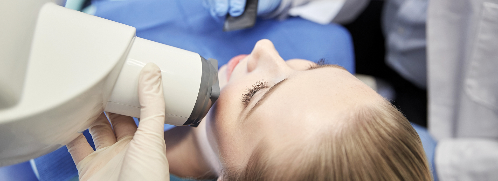
During a digital radiograph, a compact electronic sensor is positioned comfortably in the mouth or a small external device is aligned for extraoral images. The X-ray is taken and the resulting image appears on a computer monitor in seconds, allowing the dentist to review it with the patient immediately. Protective measures such as a lead apron and thyroid collar are used when indicated to further reduce exposure.
Capture time is brief and most patients experience little to no discomfort from the sensor placement. Staff will explain what the images show and how they relate to any recommended care so patients understand findings and next steps. If additional views are needed, clinicians can often adjust contrast or magnification before considering another exposure.
Once captured, digital radiographs are saved in the patient’s electronic health record where they are organized chronologically and protected by the practice’s security measures. Access controls, encrypted storage, and regular backups help maintain confidentiality and ensure images are available for future visits. Electronic storage removes the need for physical film archives and makes retrieval faster for both routine checks and specialty consultations.
When coordination with a specialist or referral is required, images can be exported in standardized formats that preserve diagnostic detail. Secure transfer methods and proper authorization procedures ensure that records are shared only with the appropriate clinicians. This streamlined sharing helps maintain continuity of care while protecting patient privacy.
Digital imaging provides tools such as contrast adjustment, magnification, and measurement overlays that assist clinicians in detecting early decay, hairline fractures, bone loss, and other subtle conditions. These enhancement features increase confidence in diagnosis and help clinicians document findings precisely in the chart. Improved visualization supports clearer communication with patients about recommended treatments and expected outcomes.
High-quality digital images also facilitate interdisciplinary planning by providing consistent, reproducible images that specialists can review alongside clinical exams. For restorative work, implant placement, or orthodontic planning, radiographic data can be integrated with other records to design solutions tailored to a patient’s anatomy. The result is more predictable treatment sequencing and better-informed clinical decisions.
Digital radiography generally uses lower doses of radiation than film X-rays, which makes it a safer option for patients who require imaging at shorter intervals. For children, clinicians use size-appropriate sensors and child-focused exposure settings to minimize dose while maintaining diagnostic value. Imaging for pregnant patients is limited to situations where it is clinically necessary and is performed with appropriate shielding and adherence to established guidelines.
Decision-making always balances diagnostic need and patient safety, and clinicians will avoid unnecessary exposures while ensuring that essential information is obtained. When imaging is required, modern digital systems combined with careful technique provide diagnostic images with attention to minimizing radiation. Patients are encouraged to discuss any concerns about imaging with their dental team so care can be individualized.
Digital radiographs are useful for identifying a wide range of conditions, including dental decay between teeth, root and periapical pathology, bone loss from periodontal disease, and fractures in tooth structure. They are also valuable for assessing the fit and condition of restorations, evaluating implant sites, and monitoring the development and position of teeth. The ability to compare current and prior images helps clinicians detect gradual changes over time.
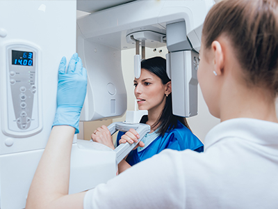
Specialized digital techniques, such as panoramic or cone-beam imaging when indicated, can reveal broader anatomical information for surgical planning and complex diagnoses. The diagnostic scope depends on the type of image taken and the clinical question being addressed. Clinicians select the appropriate imaging modality to provide the information needed for safe, effective care.
Because digital images are standardized and easily shared, general dentists, orthodontists, periodontists, and oral surgeons can review the same high-quality images when planning care. Electronic transfer of images reduces delays associated with physical film and allows specialists to assess findings before or during consultations. Shared access to the same visual data helps align treatment objectives and timelines across disciplines.
Simultaneous review of images also makes it easier to discuss complex cases with patients and among clinicians, improving clarity about recommended steps and expected outcomes. When additional input is needed, images can be annotated or supplemented with measurements to support collaborative decision-making. This coordinated approach helps reduce unnecessary procedures and supports efficient, cohesive care plans.
Digital radiography removes the need for film processing chemicals, physical storage of film, and many paper-based workflows, which reduces the environmental footprint of imaging procedures. Eliminating chemical developers and film disposal helps practices operate more sustainably and comply with environmental safety standards. Digital files also reduce the space and labor associated with archiving and retrieving photographic film.
From an operational standpoint, immediate access to images speeds appointment flow and reduces administrative steps tied to film handling. Staff can pull prior images quickly, include visuals in patient education during visits, and exchange records with specialists electronically. These efficiencies contribute to smoother scheduling, clearer communication, and better use of clinical time.
If you have questions about how digital radiography is used during your visit or about safety and imaging protocols, our team is available to explain the process and what to expect. Towne Dental & Orthodontics in Waller, TX, can describe the specific sensors and software we use, how images are stored, and the protections in place to minimize exposure. Talking with clinical staff before imaging helps patients understand the clinical rationale and feel comfortable with the procedure.
Patients who want more detailed information can request an explanation during their appointment or ask for educational materials that outline imaging steps and safety measures. The dental team can review any prior images, show comparisons, and explain how radiographs inform diagnosis and treatment planning. Open communication ensures patients are informed partners in their care.