Our Office
Visit Us Online

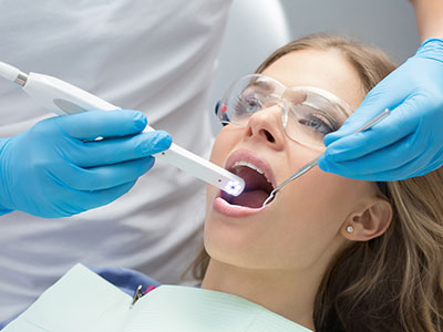
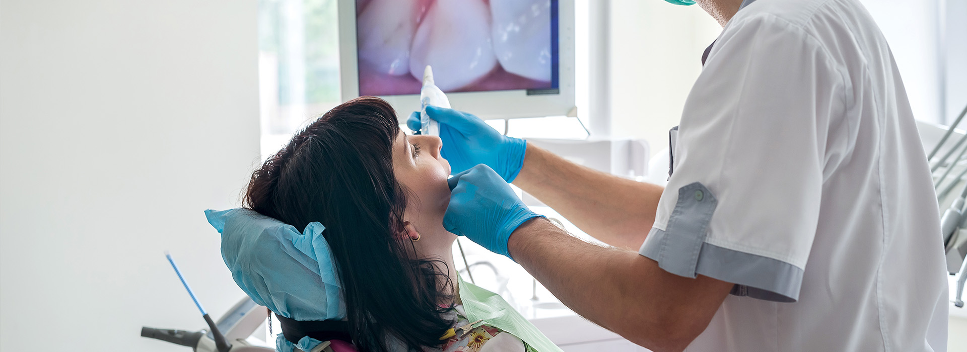
An intraoral camera is a compact, pen-sized imaging device designed to capture high-resolution color images from inside the mouth. With a small lens and built-in lighting, it can reach areas that are difficult to see with the naked eye or a standard mirror. The live feed from the camera displays on a monitor in real time, giving patients and clinicians an enlarged, accurate view of teeth, gums, and other soft tissues.
Unlike traditional visual exams, intraoral imaging provides crisp detail, which helps clinicians identify tiny cracks, early decay, and subtle changes in the soft tissues. The camera’s close-up perspective improves diagnostic accuracy by revealing surface texture, margins of restorations, and interproximal spaces. Because the images are full color and high resolution, they make it easier to differentiate healthy tissue from areas that may need attention.
Beyond still photos, many intraoral camera systems record short video clips and support image enhancement tools such as contrast adjustment and magnification. These features help the dental team document findings consistently and track changes over time. When used as part of a comprehensive exam, the intraoral camera becomes a valuable extension of the clinician’s senses, improving both assessment and patient communication.
When the intraoral camera is used during an appointment, patients can see exactly what the clinician sees on a monitor. This shared visual reference transforms a routine exam into a collaborative discussion: patients view problem areas up close and receive explanations grounded in visible evidence. That transparency helps people understand the condition of their oral health and the reasoning behind recommended care.
Seeing an enlarged image of a tooth or gum issue often clarifies why a clinician recommends a particular treatment, which can reduce uncertainty and build confidence in the plan. Visual documentation also helps patients recognize subtle oral hygiene challenges, like missed plaque along the gumline or early enamel erosion, making home care instructions more actionable and easier to follow.
For patients who have anxiety about dental care, the camera can be reassuring because it demystifies the process. Instead of relying solely on verbal descriptions, patients receive a concrete visual record of their mouth’s condition, which supports informed decisions and encourages active participation in their care.
Intraoral cameras have practical applications across many aspects of general and restorative dentistry. They are commonly used to document cavities, evaluate margins of crowns and fillings, and screen for oral lesions that warrant further evaluation. Because the device can reveal subtle anatomical details, clinicians often discover issues in early stages when treatments can be more conservative and less invasive.
During restorative procedures, intraoral images help verify the fit and finish of crowns, bridges, and other prosthetics before final cementation. For preventive care, they serve as an educational tool to illustrate how plaque accumulates or how tooth wear progresses. In specialty workflows—such as periodontal monitoring or endodontic assessment—high-resolution images complement radiographs and tactile examination, creating a fuller clinical picture.
Images captured with the camera can be exported and shared with other members of the dental team, specialists, or dental laboratories as needed. Because the files are precise and portable, they improve coordination of care and support consistent decision-making when multiple providers are involved in a patient’s treatment.
Intraoral images are routinely saved as part of the patient’s chart and become a permanent component of the clinical record. Keeping visual documentation enables the dental team to compare current findings with prior images, which is especially useful for monitoring progression or healing. This longitudinal view aids in early detection of changes and helps tailor follow-up intervals and care plans.
Stored images also streamline communication with laboratories and specialists by providing exact visual references that describe the intraoral situation. A well-documented image can reduce misinterpretation and speed up collaborative workflows, from shade selection to prosthetic design. Digital records are securely maintained according to office protocols, ensuring they remain accessible for treatment planning and review.
Because intraoral imaging is integrated into modern practice management systems, clinicians can present findings alongside radiographs, treatment notes, and care recommendations during a visit. This integrated approach supports clearer documentation and helps ensure that clinical decisions are well-supported by objective visual evidence.
Intraoral cameras are engineered for patient comfort and safety. The devices are lightweight and designed to navigate the mouth gently, with smooth surfaces that are easy to sterilize or protect with disposable sleeves. The examination process is noninvasive and typically adds only a few moments to a standard dental visit while delivering valuable diagnostic information.
Modern camera systems emphasize image quality through advanced lenses, LED lighting, and sensor technology that reduces glare and color distortion. These improvements result in clear, true-to-life photos that clinicians can rely on for precise assessment. Many systems also include software that enables measurements and annotations, which are useful for documenting lesion sizes or tracking restorative margins.
Practices that adopt intraoral imaging do so to enhance patient safety and diagnostic confidence, not to replace standard exams or radiographs. When paired with a thorough clinical evaluation and appropriate imaging studies, intraoral cameras contribute to more informed diagnoses and well-documented care plans.
Summary: Intraoral cameras bring clarity and collaboration to dental care by delivering detailed, real-time images of the mouth. They improve diagnosis, support treatment planning, and help patients better understand their oral health. At Towne Dental & Orthodontics, we incorporate this technology into routine exams to enhance communication and documentation. Contact us for more information about how intraoral imaging can support your dental care.
An intraoral camera is a small, handheld imaging device with a miniaturized lens and integrated lighting that captures high-resolution color images from inside the mouth. The device transmits a live feed to a chairside monitor so clinicians and patients can view enlarged views of teeth, gums, and soft tissues in real time. Many systems record still photos and short video clips that can be saved to a digital chart for later review.
The camera’s close-up perspective reveals surface texture, margins of restorations, and interproximal spaces that are difficult to see with the naked eye. Built-in software often includes magnification, contrast adjustment, and annotation tools that enhance diagnostic clarity. These features make the intraoral camera a practical extension of the clinical exam rather than a replacement for other diagnostic tools.
An intraoral camera provides crisp, full-color images that help clinicians detect subtle changes such as hairline cracks, early enamel breakdown, and localized soft-tissue abnormalities. The magnified view clarifies surface details and restorative margins, which supports earlier and more conservative treatment when appropriate. Combining these images with tactile exam and radiographs creates a fuller clinical picture.
Because images are recorded and compared over time, clinicians can monitor progression or healing with objective visual evidence. Software measurements and annotations allow for consistent documentation of lesion size or restoration fit. This longitudinal record enhances decision-making and follow-up planning.
No, intraoral cameras do not replace dental radiographs; they complement them by providing detailed surface and soft-tissue images that x-rays cannot show. Radiographs reveal internal structures, bone levels, and interproximal decay beneath the surfaces, while intraoral cameras highlight visual surface changes and soft-tissue concerns. Together, the two modalities provide a more complete diagnostic assessment.
Clinicians integrate camera images and radiographs during exams to correlate findings and confirm treatment needs. The camera can guide targeted radiographic imaging by highlighting suspicious areas that warrant further evaluation. Using both tools helps ensure diagnoses are accurate and well-documented.
Showing patients an enlarged image of a problem area turns abstract descriptions into concrete visual evidence, which supports clearer conversations about treatment options. When patients can see plaque accumulation, early decay, or a worn restoration up close, they better understand the rationale behind clinical recommendations. This transparency helps patients make informed decisions about their care.
Images captured during the visit can also be annotated to illustrate specific concerns and saved to the patient’s record for later review. Clinicians can use these visuals to explain home-care priorities and measurable goals, improving patient engagement and adherence to recommendations. The result is more collaborative, evidence-based treatment planning.
Yes, intraoral imaging is noninvasive and designed for patient comfort; cameras are lightweight with smooth contours so they glide easily inside the mouth. Examinations typically add only a few moments to a standard visit and do not produce radiation exposure because they capture visible light images rather than x-rays. Disposable sleeves or sterilization protocols keep the device hygienic between uses.
The real-time visual feedback is often reassuring to anxious patients because it demystifies clinical findings and reduces uncertainty. For children and sensitive patients, clinicians can take extra care to make the process gentle and explain each step. Overall, intraoral imaging enhances comfort and communication without adding risk.
Intraoral images are routinely saved as part of the digital patient chart and become permanent clinical documentation used for monitoring and treatment planning. Modern practice management systems store these files alongside radiographs and clinical notes, enabling clinicians to present a cohesive record during follow-up visits. Access to images is controlled by office protocols and user permissions to protect patient privacy.
Digital images are maintained according to HIPAA and practice-specific security measures, which can include encrypted storage, secure backups, and restricted user access. When images are shared with specialists or laboratories, that exchange is performed through secure channels to maintain confidentiality. Patients can request copies of their images for continuity of care when needed.
An intraoral camera is effective at revealing early surface-level problems such as small carious lesions, enamel erosion, hairline fractures, and marginal breakdown around fillings or crowns. It also helps identify soft-tissue changes like ulcers, recession, or suspicious lesions that may need further evaluation. Detecting these issues early often allows for less-invasive treatment options.
In preventive care settings, the camera documents plaque accumulation patterns and areas missed by daily brushing, which can inform personalized home-care instruction. For patients undergoing restorative or periodontal therapy, serial images help track healing and treatment response. Early visual detection supports proactive, conservative care.
During restorative work, intraoral images help clinicians verify the fit, margin integrity, and finish of crowns, bridges, and fillings before final cementation. The magnified views make it easier to confirm that restoration contours align with adjacent teeth and that margins are flush and polished. Recording images at key stages documents the quality of work and supports quality assurance.
Camera images also facilitate communication with dental laboratories by providing exact visual references for shade selection and prosthetic design. When adjustments are required, annotated images clarify the issue and reduce the likelihood of misinterpretation. This coordination improves efficiency and outcomes for complex restorative cases.
Yes, intraoral images are digital files that can be exported and shared with specialists or dental laboratories to support collaborative care. Sharing precise visual references improves treatment coordination by giving consultants and technicians a clear view of the intraoral situation. Many practices transmit images through secure portals or encrypted channels to protect patient privacy.
Providing images alongside radiographs and clinical notes reduces ambiguity in referrals and lab prescriptions, which can speed up case planning and improve the accuracy of prosthetic work. Shared images also make multidisciplinary discussions more productive by aligning all providers around the same visual evidence. Properly documented images ensure continuity and clarity throughout the treatment process.
Towne Dental & Orthodontics incorporates intraoral imaging into routine exams to enhance patient communication and clinical documentation. Using the camera in our Waller, TX office allows clinicians to show patients enlarged views of concern areas and to record objective images that support diagnosis and follow-up care. Integrating these images with radiographs and clinical notes creates a comprehensive digital record for each patient.
When appropriate, clinicians use intraoral photos to guide preventive instruction, verify restorative work, and coordinate care with specialists or laboratories. Patients who want to review their images or discuss what was documented during a visit are encouraged to ask questions so the team can explain findings and recommended next steps. This approach helps ensure transparent, well-supported treatment planning.